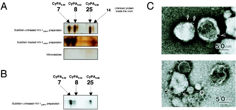
FIG. 4.
2D gel image analysis, Western immunoblot analysis with anti-CyPA antibody, and immunoelectron microscopic analysis. (A) After 2D gel electrophoresis and protein visualization by silver staining, the 2D gel images of the non-subtilisin-treated HIV-1LAV-1 preparation, the subtilisin-treated HIV-1LAV-1 preparation, and microvesicles alone were expanded. The expanded image of the non-subtilisin-treated HIV-1LAV-1 preparation shows four spots (spots 7, 8, 14, and 25). Spot 25 completely disappeared on subtilisin treatment. This result suggests that CyPA6.88 exists on the viral surface. Spots 7, 8, 14, and 25 were not detected in the image of the microvesicles. The spot numbers are in agreement with those in Fig. 2. (B) Spots 7, 8, and 25 on the 2D gel of the non-subtilisin-treated HIV-1LAV-1 preparation were subjected to Western immunoblot analysis with anti-CyPA antibody. (C) A microvesicle-contaminated HIV-1 preparation was fixed with 0.1 M sodium phosphate buffer (pH 7.4) containing 4% paraformaldehyde and 0.25% glutaraldehyde at 4°C. After brief washing, the samples were mounted on carbon-coated nickel grids. Double immunolabeling techniques were used as follows. The first step was rabbit anti-CyPA antibody (Upstate Biotechnology) and then 15-nm-diameter gold-labeled secondary antibody, and the second step was murine anti-gp120 (strain IIIB) monoclonal antibody (ImmunoDiagnostics, Inc., Woburn, Mass.) and then 5-nm-diameter gold-labeled secondary antibody. The immunolabeled samples were negatively stained with 3% uranyl acetate and examined with a transmission electron microscope. The asterisk indicates a microvesicle; arrows and arrowheads indicate gp120 and CyPA, respectively.

