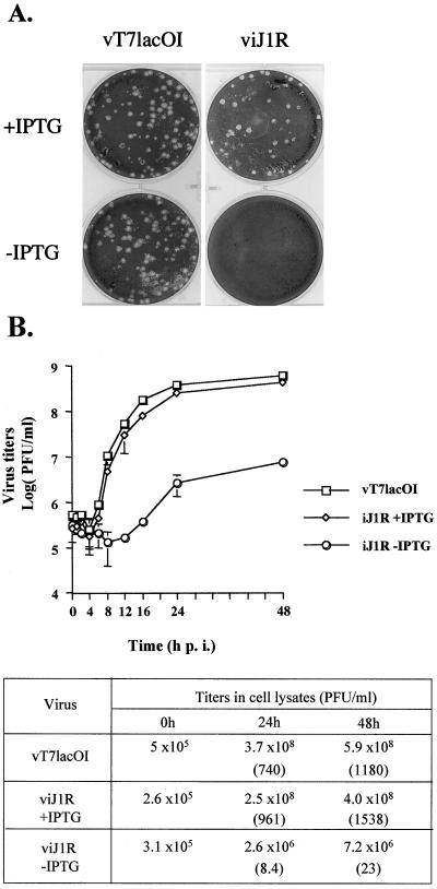
FIG. 4.
Characterization of viJ1R. (A) viJ1R mutant virus does not form plaques on BSC40 cells. BSC40 cells were infected with vT7lacOI or viJ1R, incubated in medium for 3 days, fixed, stained with crystal violet, and photographed. (B) One-step growth curve analysis of viJ1R virus. BSC40 cells were infected with parental vT7lacOI or viJ1R at an MOI of 5 PFU per cell; incubated in normal medium or medium with IPTG; and harvested at 0, 1, 2, 4, 6, 8, 12, 16, 24, and 48 h p.i. Error bars indicate standard deviations. Virus titers in the lysates were determined by plaque formation assays with BSC40 cells and are listed in the table. Numbers in parentheses are the fold increase in virus titer, determined as the virus titer at 24 or 48 h p.i. divided by the virus titer at 0 h.

