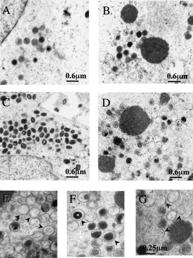
FIG. 6.
Electron micrographs of vaccinia virion morphogenesis in cells infected with viJ1R. BSC40 cells were infected with viJ1R virus at an MOI of 20 PFU per cell either in the presence (A and C) or in the absence (B and D to G) of IPTG and fixed at 12 h (A and B) or 24 h (C to G) p.i. for electron microscopy. Photos were taken at magnifications of ×12,000 (A to D) and ×30,000 (E to G). Arrowheads in panels E to G represent aberrant membrane structures, such as empty IV (E), double-layer membranes (F), and half-circle membranes (G).

