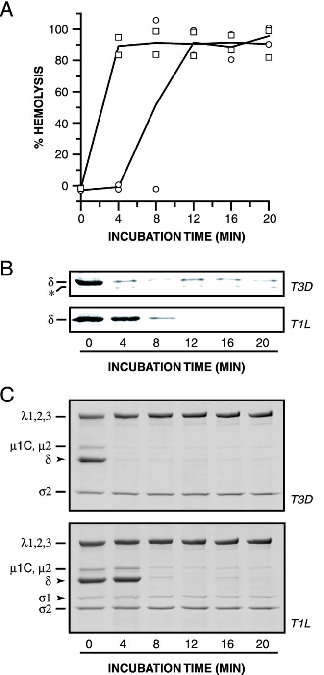
FIG. 3.
Effect of Cs+ ions and RBCs on hemolysis and the conformational state of μ1. (A and B) Nonpurified ISVPs of strains T1L (circles) or T3D (squares) (4 × 10¹² particles/ml) were incubated with RBCs in reaction buffer (50 mM Tris-Cl [pH 7.5]) containing CsCl (300 mM) for different times at 32°C. Measurement of the amount of hemolysis (A) and assessment of the protease sensitivity of δ (B) were carried out as described for Fig. 2A and B. Results from two trials are shown in panel A. (C) Samples were generated as described for panels A and B except that no RBCs were added. Samples were treated with trypsin for 30 min on ice, and digestion was stopped by addition of soybean trypsin inhibitor. Samples were subjected to SDS-PAGE, and the viral proteins were visualized by Coomassie staining.

