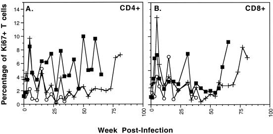
FIG. 6.
Longitudinal analysis of SIVmac239-infected macaques RM1 (○), RM2 (▪), and RM3 (+) to assess expression of the Ki67 antigen as an indicator of cellular proliferation. The percentage of cells expressing Ki67 in the peripheral blood within CD4+ cells (A) and CD8+ cells (B) is depicted for each macaque. Ki67+ T-cell levels preinfection ranged from 1 to 4% in the CD4+ cells and 1 to 3% in the CD8+ cells.

