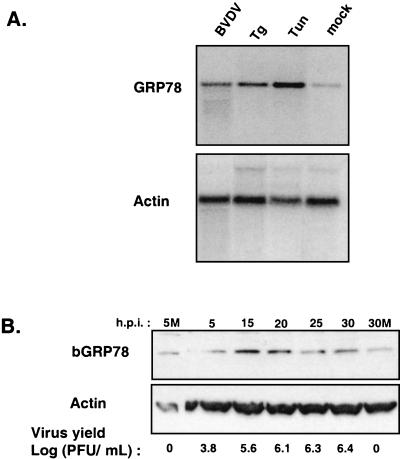
FIG. 2.
Induction of GRP78 expression by BVDV infection and ER stress-inducing agents. (A) MDBK cells were infected with BVDV or treated with either 1 μg of tunicamycin (Tun) per ml or 50 nM thapsigargin (Tg) and harvested at 24 h postinfection (h.p.i.). Northern blot analysis was performed with a radiolabeled bovine GRP78 cDNA probe. The blot was stripped and rehybridized to a radiolabeled actin cDNA probe. (B) Immunoblot analysis of GRP78 protein levels during a time course of BVDV infection. MDBK cells were infected with BVDV and harvested at the indicated times. Approximately 100 μg of total cell protein from cells harvested at each time point was fractionated by SDS-PAGE on 4 to 20% polyacrylamide gels and subjected to immunoblot analysis with anti-KDEL polyclonal antibodies and monoclonal antibodies against actin. Enhanced chemiluminescence was used to visualize immune complexes. Virus yields in the culture supernatants were measured by plaque assay. 5M and 30M, 5 and 30 h post-mock infection, respectively.

