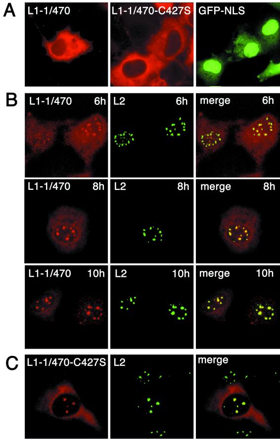
FIG. 3.
Cellular localization of mutant L1 and wt L2 proteins. (A) COS-7 cells were infected for 10 h with recombinant vaccinia virus encoding mutant L1-1/470 or L1-1/470-C427S or were transfected with pEGFPGFP-NLS coding for the HPV33 L1-NLS fused to the carboxy terminus of dimeric GFP (GFP-NLS). Capsid proteins were visualized after 10 h by immunofluorescence. GFP was visualized 40 h after transfection. (B) COS-7 cells were coinfected with recombinant vaccinia viruses encoding wt L2 and mutant L1-1/470, respectively. Capsid proteins were visualized by immunofluorescence at the indicated times after infection. (C) COS-7 cells were infected with recombinant viruses encoding wt L2 and mutant L1-1/470-C427S, respectively. Cells were fixed 8 h after infection, and the capsid proteins were visualized by immunostaining.

