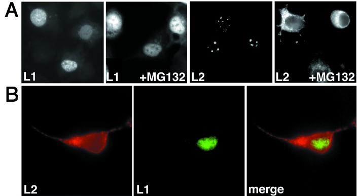
FIG. 5.
Localization of wt L1 and L2 in cells treated with MG132. HuTK− cells were coinfected with vaccinia viruses encoding wt L1 and L2, respectively. MG132 was added 3 h after infection, and the capsid proteins were visualized 6 h later using immunofluorescence. The cells were infected with one virus (A) or two viruses (B). (A) The nuclear dots lacking L1, as seen in the L1 +MG132 panel, represent nucleoli.

