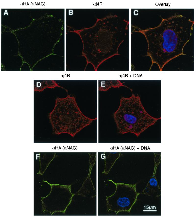
FIG. 7.
Localization of j4R and αNAC in cells. Cells were infected with the MVA T7 strain of vaccinia virus and transfected with plasmids pcDNA HA αNAC and pcDNAj4R together (A to C) or separately (D to G). At 12 h posttransfection cells were fixed and stained with anti-HA rat MAb to detect HA αNAC (green; panels A, F, and G) and anti-j4R serum (red; panels B, D, and E). DNA was stained with ToPro3 (blue; panels C, E, and G). Overlaid images of (i) panels A and B, plus DNA, are shown in panel C; (ii) panel D, plus DNA, are shown in panel E; and (iii) panel F plus DNA stain are shown in panel G. Cells were imaged by confocal microscopy, and single optical sections are shown.

