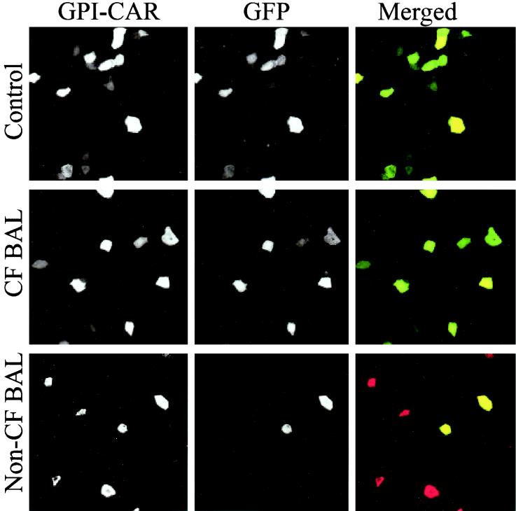
FIG. 3.
Immunostaining of human airway epithelium displaying GPI-CAR infected with Ad2/GFP. Airway epithelium displaying GPI-CAR on the apical surface was infected with 10 MOI of Ad2/GFP in the presence of medium alone, CF BAL, and non-CF BAL. Two days later, epithelia were fixed with 4% paraformaldehyde, and a mouse anti-Flag monoclonal antibody was added to the apical surface of the epithelium for 1 h at 37°C. Subsequently, these epithelia were incubated with donkey anti-mouse IgG conjugated with Texas Red fluorophore. Apical staining for GPI-CAR and GFP expression was evaluated by laser confocal microscopy (MRC-1024; Bio-Rad) at ×60 magnification. Merged images of GPI-CAR (red) and GFP (green) are shown. Cells that express both GPI-CAR and GFP appear yellow.

