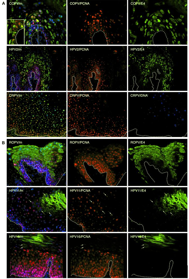
FIG.1.
Heterogeneity in the timing of events during the life cycles of human and animal papillomaviruses. (A) Tissue sections from a mucosal lesion caused by COPV (top row) and a cutaneous lesion caused by HPV2 (middle row) were double stained using antibodies to E4 (green) andPCNA (red) before being counterstained with DAPI (blue) to visualize cell nuclei. Surrogate markers of E7 expression (PCNA) were confined to sporadic cells in the lowest parabasal layers. E4 (green) was first detected in these cells as they migrated through the lower layers of the epithelium. E4 was expressed in the basal layer in lesions caused by COPV and in a few cells above the basal layer in lesions caused by HPV2. Surrogate markers of E7 were lost soon after the appearance of E4. The merged images (m) are shown at the left. A section through a cutaneous lesion caused by CRPV is shown in the bottom row after immunostaining to detect PCNA (red) and in situ hybridization to detect viral genome amplification (blue). The nuclei were counterstained using Sytox green. Surrogate markers of E7 expression (PCNA) did not persist to the epithelial surface and were lost soon after the onset of viral genome amplification (blue). Genome amplification began in the mid-spinous layers in lesions caused by CRPV. The broken lines indicate the positions of the basal layers. The images were taken using a 10× (COPV and HPV2) or 20× (COPV/m inset) objective. (B) Tissue sections from mucosal lesions caused by ROPV (top row), HPV11 (middle row), and HPV16 (bottom row) were double stained with antibodies to E4 (green) and PCNA (red) before being counterstained with DAPI (blue) to visualize cell nuclei. The merged images are shown at the left. In such lesions, surrogate markers of E7 expression (PCNA; red) usually persisted into the upper epithelial layers but were lost following the expression of E4. E4 was rarely detected in the lower epithelial layers in lesions caused by HPV11 and -16. Cells expressing both E4 and PCNA are indicated by arrows in the HPV11 and HPV16 images. The broken lines indicate the positions of the basal layers. The images were taken using a 20× objective.

