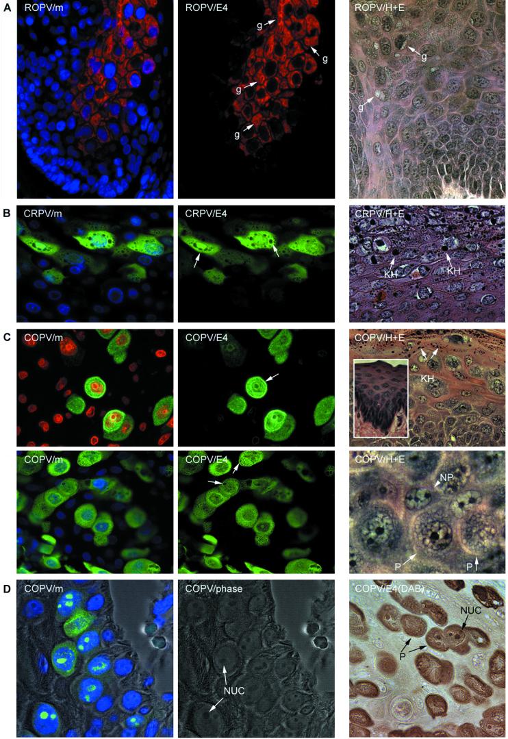
FIG.5.
Intracellular distribution of the E4 proteins of animal papillomaviruses. (A) The E4 protein of ROPV (red) is predominantly cytoplasmic and is associated with inclusion granules (center image, g) similar to those seen in cutaneous lesions caused by HPV1. The nuclei werecounterstained with DAPI (blue) and are visible in the merged image at the left (ROPV/m). The E4 granules (g) are also shown in the hematoxylin- and eosin-stained image at the right (labeled ROPV/H+E). The images were taken using a 40× objective. (B) The E4 protein of CRPV (green) is distributed throughout the nucleus and the cytoplasm. The nuclei were counterstained with DAPI (blue) and are visible in the merged image shown on the left (CRPV/m). The cytoplasmic structures that did not stain with antibodies to E4 are keratohyalin granules (center image, arrows). They are also shown (KH) in the hematoxylin- and eosin-stained image (CRPV/H+E) on the right. (C) The E4 protein of COPV (green) was cytoplasmic and nuclear but was also associated with the nuclear and cellular periphery (upper center image, arrow). The nuclei were counterstained with either propidium iodide (upper left image, red) or DAPI (lower left image, blue). A granular pattern was apparent in the cytoplasm of some cells (granule-like structures [arrows] in lower center image). Keratohyalin granules (KH) are abundant in COPV-induced warts and are shown in the hematoxylin- and eosin-stained image (COPV/H+E; upper right). The surrounding mucosal epithelium is devoid of keratohyalin (inset). Cells expressing COPV E4 had a characteristic morphology that may result from the presence of E4 inclusion granules. The permissive cells (P) that express E4 and the nonpermissive cells (NP) that do not express E4 are shown in the hematoxylin- and eosin-stained image on the lower right. (D) In the lower epithelial layers of experimental warts caused by COPV, nuclear E4 protein (green) was found associated with the nucleoli. At the left (COPV/m), the E4 and DAPI (blue) stain is shown as an overlay of the phase-contrast image. The phase-contrast image is shown in the center panel to indicate the presence of the nucleoli (NUC). The presence of the permissive “granular” cells (P) and the nucleoli is clearly visualized in the immunoperoxidase stain (brown) shown on the right [COPV/E4 (DAB)].

