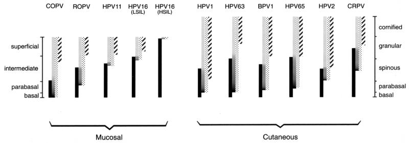
FIG. 8.
The regulation of early and late events in lesions caused by different papillomavirus types. The timing of life cycle events in lesions caused by animal and human papillomaviruses is indicated by the bars. The shaded bars show the presence of amplified viral DNA, while the stippled bars show the extent of E4 expression. The expression pattern of surrogate markers of E7 is shown by the solid bars, whereas the L1 expression pattern is indicated by the hatched bars. The darker region at the bottom of the shaded bars indicates the region where vegetative viral genome amplification is thought to occur. Mucosal epithelial tissue infected by COPV, ROPV, HPV11, and HPV16 is divided into four layers, shown on the left. Cutaneous tissue infected by HPV1, HPV63, BPV1, HPV65, HPV2, and CRPV is divided into five layers, shown on the right. For each group of papillomaviruses, those that initiate their late events in the lower epithelial layers are shown on the left. Viruses that initiate their late events close to the epithelial surface are shown on the right. COPV triggers late events in the lowest epithelial layers. Lesions caused by HPV11 and HPV16 (LSIL) usually support late gene expression only in the upper half of the epidermis, whereas late events are not always supported in HSIL. In all instances, the loss of surrogate markers of E7 does not occur until after E4 has accumulated to detectable levels.

