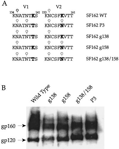
FIG. 3.
(A) Amino acid alignment of the V1 and V2 regions of wild-type, variant, and mutant envelope glycoproteins. Bold letters designate amino acid changes. ○, presence of glycosylation site. WT, wild type. (B) Immunoblot analysis of wild-type, P3 variant, and mutant envelope glycoproteins. HEK-293T cells were transfected with proviral DNAs. Forty-eight hours posttransfection, cells were harvested and lysed and proteins were separated by sodium dodecyl sulfate-4 to 12% polyacrylamide gel electrophoresis. Envelope gp120s were detected by immunoblotting with a polyclonal goat anti-gp120 antibody. Positions of gp160 and gp120 are denoted.

