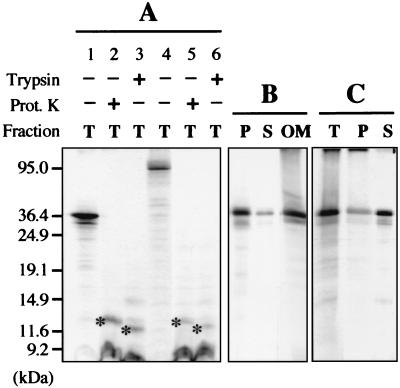
FIG. 3.
In vitro protein insertion assays in the presence of isolated plant mitochondria. (A) After the insertion assay in the presence of potato mitochondria, incorporation of the in vitro-translated CIRV 36K (lanes 1 to 3) and 95K (lanes 4 to 6) polypeptides was analyzed in the total mitochondrial fraction (T); mitochondria were either untreated (lanes 1 and 4) or treated with proteinase K (lanes 2 and 5) or trypsin (lanes 3 and 6) after insertion. The specific polypeptides protected against proteinase K or trypsin digestion in the mitochondrial fractions are indicated by an asterisk. (B) After the insertion assay in the presence of potato mitochondria, incorporation of the CIRV 36K polypeptide was analyzed in the pellet (P) and supernatant (S) fractions following carbonate extraction of the mitochondria and in the mitochondrial outer membrane fraction (OM). (C) The insertion assay was run in the presence of a potato mitochondrial outer membrane preparation instead of intact mitochondria; incorporation of the CIRV 36K polypeptide was subsequently analyzed in the membrane fraction (T) and in the pellet (P) and supernatant (S) following carbonate extraction of the membrane fraction. Samples were analyzed by conventional SDS-PAGE. In vitro-translated full-length viral proteins and truncated forms were used as molecular size markers, and their positions are indicated on the left.

