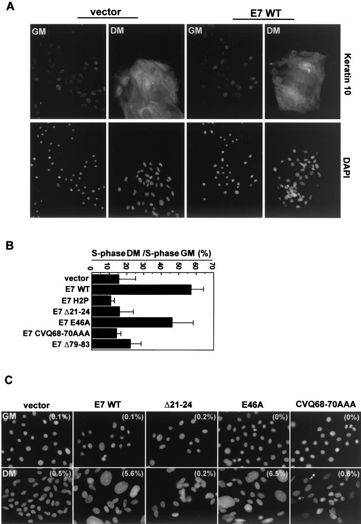
FIG.2.
Uncoupling of cell cycle arrest and differentiation by E7. (A) Keratin 10 expression in differentiated HFKs. The upper panels show keratin 10 labeling with an anti-keratin 10 antibody (DE-K10; Neomarkers), and the lower panels show nuclei counterstained with DAPI (Sigma). Images were collected with a 20× objective on a Nikon Eclipse TE300 microscope equipped with a charge-coupled-device camera (Roper Scientific) and Metamorph software (Universal Imaging Co.). GM, growth medium; DM, differentiation medium. (B) S-phase induction in differentiated HFKs. The S-phase populations are graphed normalized to populations of undifferentiated cells. Shown is a summary of the results of three experiments. Error bars represent standard deviations. (Raw data are presented in Table 1). (C) Giant nuclei in E7-expressing, differentiated HFKs. Images of DAPI-stained nuclei were collected at the same magnification with the equipment described for panel A. Examples are shown, with the percentage of cells with giant nuclei (of 300 cells counted) indicated in parentheses on each image. Nuclei were considered giant if their area was at least four times greater than the average size of nuclei in vector-expressing cells.

