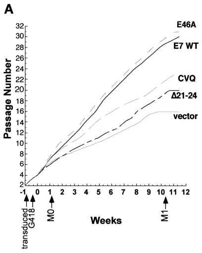
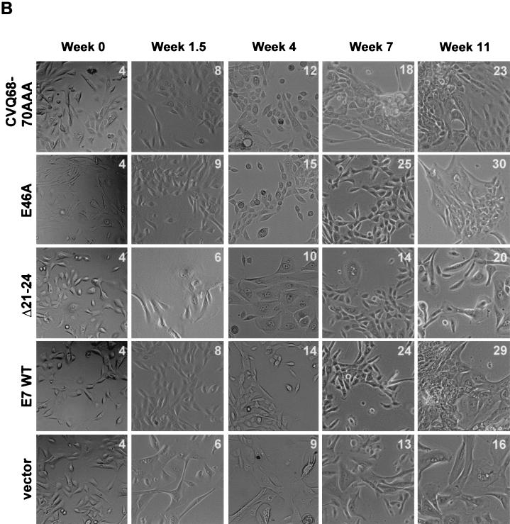
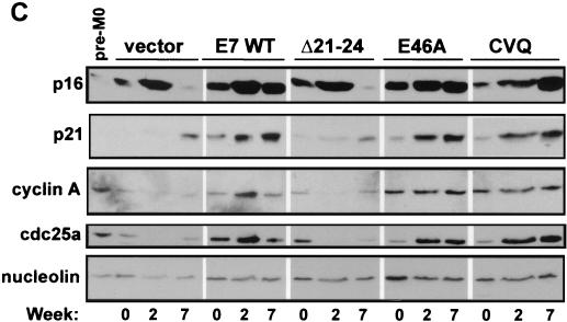
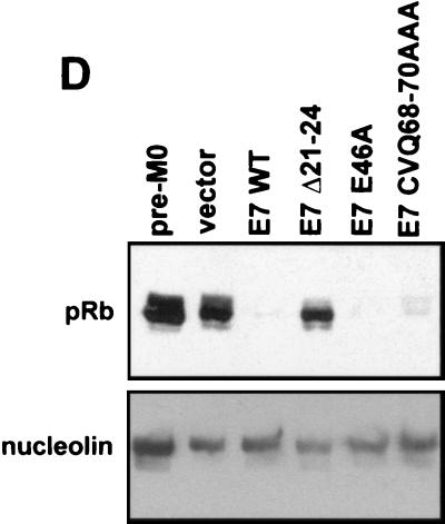
FIG. 3.
Induction of HMEC proliferation by E7. (A) HMEC passage number relative to time in culture. Times of transduction and G418 selection are indicated. The first passage after drug selection (passage 4) was designated week 0. (B) Micrographs showing HMEC morphologies at different time points. The passage number is indicated on each image. (C) Western blots probed with anti-p16 (Pharmingen), anti-p21 (Ab-2; Calbiochem), anti-cyclin A (BF-683; Pharmingen), anti-cdc25a (F-6; Santa Cruz Biotechnology), and antinucleolin (as a loading control, C23; Santa Cruz Biotechnology). (D) Western blot probed with an anti-Rb antibody (G3-245; Pharmingen) comparing Rb levels in E7-expressing HMECs with that of untransduced, pre-M0 HMECs (first lane).


