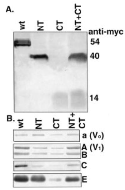
Figure 3. Levels of V-ATPase subunits in vacuoles containing the VMA13 fragments.

Vacuolar vesicles were isolated from vma13δ mutants expressing plasmid-borne copies of the intact H subunit gene (wt) or the indicated H subunit fragments (NT, CT, and NT+CT). Vacuolar proteins was solubilized, separated by SDS-PAGE, and transferred to nitrocellulose before blotting with antibodies specific for the Myc epitope on the wild-type and mutant H subunits and for different V-ATPase subunits. A, immunoblot of vacuolar vesicle protein (15 μg) probed with anti-Myc antibody. B, immunoblot of vacuolar vesicle protein probed with antibodies to Vo subunit a, V1 subunit A, V1 subunit B, V1 subunit C, and V1 subunit E. 5 μg of protein was loaded for visualization of subunits A, B, and E, and 15 μg was loaded for visualization of subunits a and C.
