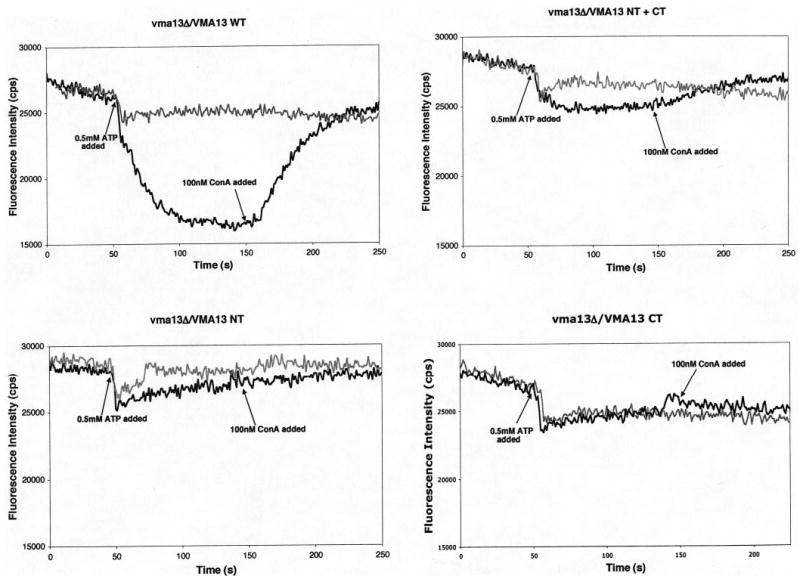
Figure 4. Proton pumping in vacuolar vesicle preparations containing intact VMA13 and VMA13 fragments.

Vacuolar vesicles (10 μg of protein per assay) isolated from the indicated strains were mixed with 1 μM ACMA in transport buffer (50 mm NaCl, 30 mm KCl, 20 mm HEPES, pH 7) in a fluorometer cuvette. Fluorescence emission intensity (black line for each plot) was monitored continuously as the mixture was stirred at 25 °C, and at the indicated point a mixture of MgSO4 and ATP was added to give final concentrations of 1 mm MgSO4 and 0.5 mm ATP (indicated as 0.5 mm ATP on each plot). After the fluorescence decrease had stabilized, 100 nm concanamycin A was added to the cuvette at the indicated time. The gray plots represent control experiments conducted for each strain in which 100 nm concanamycin A was present throughout the assay to inhibit any V-ATPase-dependent changes in fluorescence.
