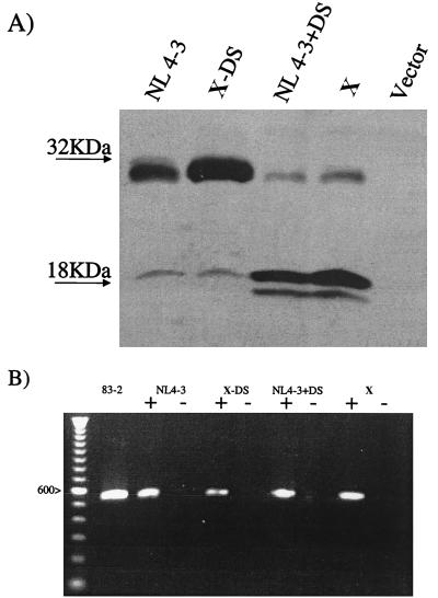
FIG. 5.
(A) Western immunoblot of strains from patient X and strains with NL4-3 or derivative Vif sequences. Cells transfected with vector sequences without Vif sequences (Vector) served as the control. The migration of molecular size markers is indicated to the left. (B) RT-PCR of strains from patient X and strains with NL4-3 or derivative Vif sequences. A plus sign indicates that the isolated RNA was reverse transcribed prior to PCR amplification, and a minus sign indicates that the isolated RNA was not reverse transcribed prior to PCR amplification. A 100-nucleotide ladder is shown on the left, and the identity of the 600-nucleotide band is indicated.

