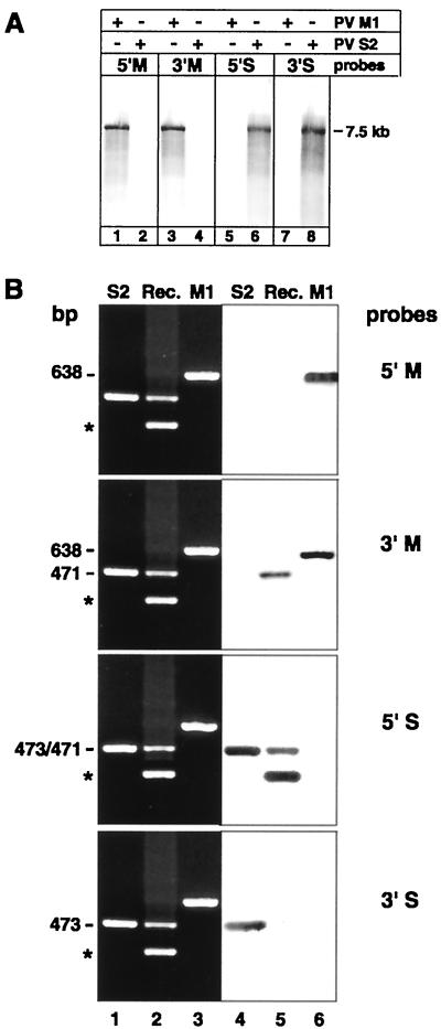
FIG. 3.
(A) Northern blot hybridization with the DIG-labeled M (lanes 1 to 4) or S (lanes 5 to 8) riboprobe on PV M1 or S2 7.5-kb-genome-length RNA extracted from corresponding virus stocks shows type specificity of probes. The position of the 7.5-kb marker RNA is indicated. +, present; −, absent. (B) Southern blot hybridization with the DIG-labeled type-specific riboprobes on cDNA from single- or double-infected cells. Ethidium bromide-stained gels (lanes 1 to 3) of RT-PCR-amplified PV S2 (lane 1), recombinant PV S2-M1 (Rec.; lane 2), and PV M1 RNA (lane 3) are juxtaposed to their corresponding blots hybridized with the DIG-labeled M and S probes (lanes 4 to 6). The probes recognize PV M1 cDNA of 638 bp or PV S2 cDNA of 473 bp at their 5′ and 3′ termini. The 471-bp cDNA of the PV S2-M1 recombinant is recognized by the 5′S and the 3′M probes but not the 5′M and the 3′S probes. The asterisks indicate nonspecific bands as in Fig. 2.

