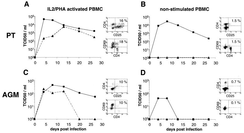
FIG. 3.
In vitro replication of SIVagm3mc and SIVagm3-X4mc. PBMCs from PT macaques (A and B) and AGM (C and D) were infected at an MOI of 0.1 with SIVagm3mc (▴) and SIVagm3-X4mc (▪). Replication was followed in IL-2-PHA-stimulated PBMCs (A and C) or nonstimulated PBMCs (B and D) by measuring the TCID50 in C8166 T cells at various time points after infection as indicated. The stimulation status of the cells was monitored by FACS analysis using a double stain for CD4-CD69 or CD4-CD25 on the day of infection. The FACS diagrams show the percentages of double-positive PBMCs (upper right boxes) for each infection.

