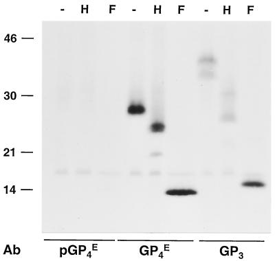
FIG. 10.
Identification of the GP4 and GP3 proteins in virions. EAV-infected BHK-21 C13 cells were labeled with [35S]cysteine from 6.5 to 11 h p.i. After removal of cell debris by low-speed centrifugation, the labeled virus present in the cell culture medium was pelleted through a cushion of 20% (wt/wt) sucrose. The pellet was then dissolved in lysis buffer containing 20 mM NEM and subjected to IP with αGP4E or αGP3 antiserum or, as a negative control, αGP4E preimmune serum (pGP4E) in the presence of 5 mM DTT. The immunoprecipitates were mock treated (−) or treated with endo H (H) or PNGase F (F). The samples were finally dissolved in LSB containing 50 mM DTT and analyzed in SDS-15% PAA gels. The values on the left are the molecular sizes, in kilodaltons, of marker proteins analyzed in the same gel. Ab, antibody.

