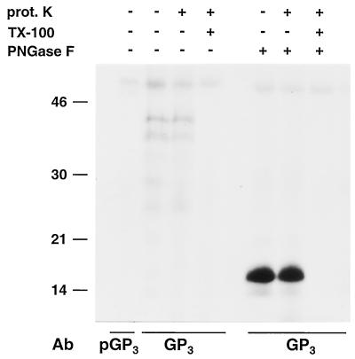
FIG. 6.
Membrane topology of the GP3 protein. ORF3 RNA transcripts were translated in vitro in the presence of canine pancreatic microsomes. Subsequently, the translation products were treated or mock treated with proteinase K (prot. K) in the absence of detergent or after disruption of the microsomal membranes with TX-100. After a 1-h incubation at 4°C, the proteinase K was inactivated by protease inhibitors. Next, the samples were immunoprecipitated with a GP3-specific antipeptide serum (GP3) or its preimmune serum (pGP3). The resulting immune complexes were each split into two equal portions that were treated or mock treated with PNGase F. The samples were dissolved in LSB containing 5% β-mercaptoethanol and analyzed in SDS-15% PAA gels. The values on the left are the molecular sizes, in kilodaltons, of marker proteins analyzed in the same gel. Ab, antibody.

