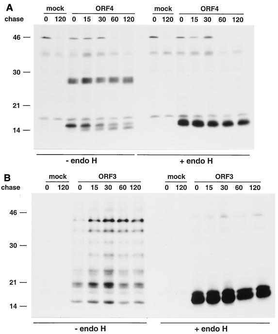
FIG. 7.
Kinetics of endo H resistance acquisition of individually expressed GP4 and GP3 proteins. BHK-21 C13 and BSR T7/5 cells were infected with vTF7.3 and transfected or mock transfected with ORF4-specific plasmid pMRI14 (A) or ORF3-encoding plasmid pAVI13 (B). The cells were pulse-labeled for 15 min with [35S]cysteine at 4.5 h p.i. and chased in the presence of 0.5 mM cycloheximide for the times indicated (in minutes). The GP4 and GP3 proteins were then immunoprecipitated from cell lysates with the αGP4E and αGP3 antisera, respectively, in the presence of 5 mM DTT. The immunoprecipitates were treated with endo H (+) or mock treated (−). The samples were finally dissolved in LSB containing 50 mM DTT and analyzed in SDS-15% PAA gels. The values on the left are the molecular sizes, in kilodaltons, of marker proteins analyzed in the same gels.

