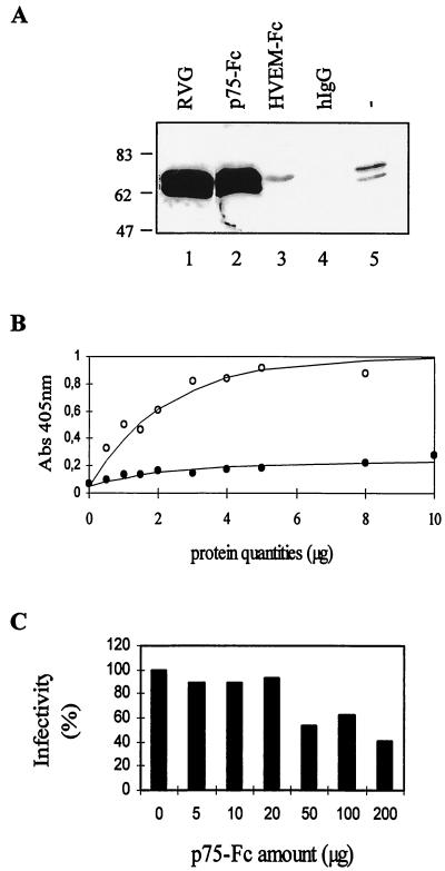
FIG. 2.
p75-Fc interacts with RVG. (A) Coprecipitation. RVG, solubilized from CVS particles with 1% CHAPS, was incubated with preadsorbed p75-Fc (lane 2), HVEM-Fc (lane 3), human IgG (lane 4), or protein A-Sepharose (lane 5). The immune complexes were analyzed by SDS-PAGE, and the presence of RVG was detected by Western blotting with anti-RVG 17 D2. In lane 1, solubilized RVG was loaded. (B) ELISA. Various amounts of purified p75-Fc (open circles) or human IgG1 (solid circles) were applied to a 96-well plate that had been coated with 200 ng of purified virus per well. Bound recombinant proteins were detected with an antihuman IgG alkaline phosphatase-conjugated antibody, with p-nitrophenyl phosphate as a substrate. A405 was measured. The data are the means from duplicate wells. (C) Neutralization. CVS (103 PFU) was incubated with different amounts of purified p75-Fc. A plaque assay was then performed with BSR cell monolayers. The residual infectivity was calculated as the ratio of virus titers obtained in the presence and absence of p75-Fc.

