
FIGURE 5.
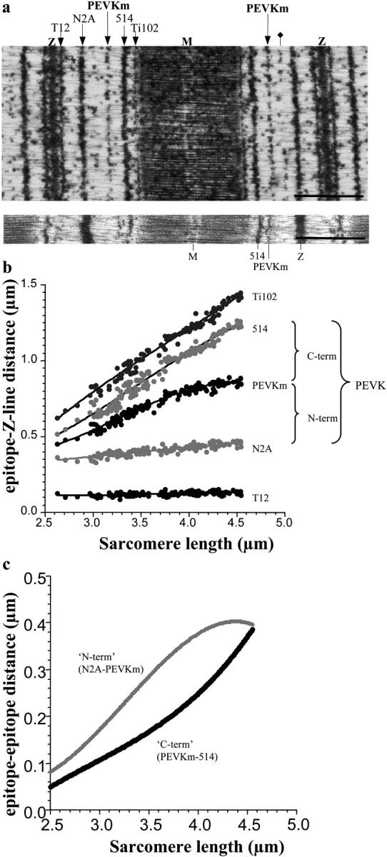
IEM of soleus muscle using different anti-titin antibodies. (a, upper panel) Immunoelectron microscopic image of a soleus muscle sarcomere stretched to an SL of 3.8 μm. Scale bar 1 μm. The fiber was labeled with a cocktail of T12, N2A, 514, and Ti102 antibodies (indicated above the figure). PEVKm indicates the extra epitope labeled with the 514 antibody. The additional 514-labeled weak epitope is labeled with a diamond. (Lower panel) Immunoelectron microscopic image of a soleus muscle sarcomere labeled with the 514 antibody only. Scale bar 1 μm. (b) Z-line to epitope distance as a function of SL. (c) Segmental extensibility within the PEVK domain (as deduced from the epitope to epitope distances as indicated) as a function of SL.