
FIGURE 6.
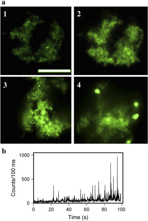
Generation of hybrid nanoclusters in situ. (a) Epifluorescence microscopy images of amyloid fibrils prestained with ThT (10 μM) followed by in situ photoreduction with different concentrations of AgNO3: 0.1 μM at time zero (panel 1) and after 2 min of irradiation at 475 nm (panel 2), 1 μM AgNO3 after 2 min of irradiation (panel 3), and 10 μM AgNO3 after 2 min of irradiation (panel 4); scale bar = 10 μm. Exposure time for collecting images was 0.08 s for panels 1 and 2 and 0.02 s for panels 3 and 4. (b) Emission blinking observed from randomly selected single Ag-ThT nanoclusters monitored during formation of nanoclusters in situ. Emission was acquired for 1000 frames with 100-ms time resolution for each frame. Fluorescence was recorded with an irradiance of 500 W/cm2.