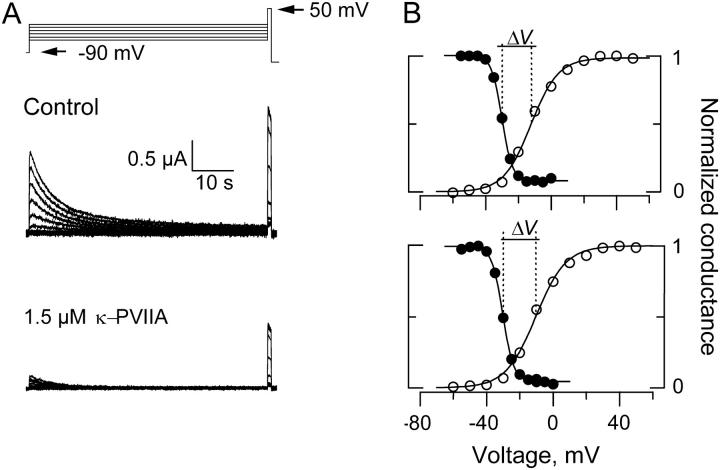
FIGURE 5.
Voltage dependent availability of Shaker-IR K-channels in the presence and absence of 1.5 μM κ−PVIIA. (A) With TEVC, 1-min conditioning pulses ranging from −60 to 0 mV separated by increments of 5 mV were applied before a 1 s test pulse to +50 mV. (B) Plots of the current in response to the test pulse voltage. Current amplitudes were normalized to the maximum current elicited at each experimental condition. Voltage dependent availability is plotted in filled circles for control (top) and in the presence of 1.5 μM κ−PVIIA (bottom). Open circles plot the relative oocyte conductance before the control voltage excursion (top) and after the voltage excursion with toxin was taken (see Materials and Methods). To calculate the conductance, we assumed a reversal potential for K+ as −100 mV and a maximal conductance of 0.015 mS. Continuous lines on top of the experimental estimates are fits to a Boltzman function of the form Y = 1/(1 + e(V−Vo)/kv). For the voltage dependent availability, the fits parameters of this experiments were Vo = −30 mV, kv = 3.1 mV−1 for the control measurements and Vo = −30.5 mV, kv = 3.3 mV−1 for the measurements in the toxin presence. For the normalized conductances, the fit parameters were Vo = −12 mV and kv = −8.3 mV−1 before the control experimental run and Vo = −10 mV and kv = −9.0 mV−1 after the toxin run. These estimations gave values of ΔV = −18 mV and ΔV = −20.1 for the control and toxin run, respectively.

