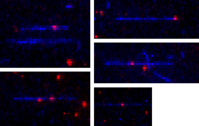
FIGURE 1.
Colocalization of actin filaments with sparsely labeled myosin. Actin filaments labeled with AEDANS (blue) at Cys374 are decorated with HMM or S1. Labeled and unlabeled myosin fragments are mixed ∼1:1000 so individual rhodamine (red) molecules can be distinguished along the length of the actin filaments. When the decorated filaments are added in the presence of excess free HMM or S1, respectively, to a poly-L-lysine coated flow chamber, the filaments are aligned well, as pictured here, due to a balance between flow forces and attachment frequency.

