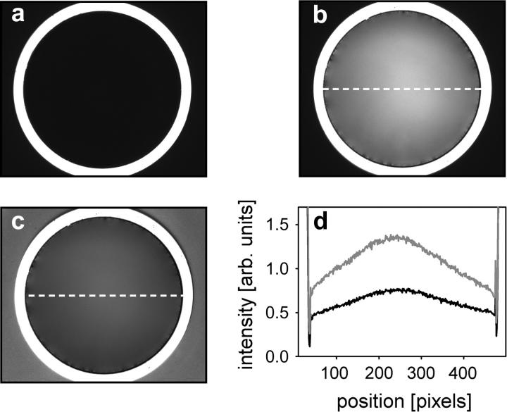
FIGURE 2.
Microscopic views of the inverted interface. (a) Appearance of the aperture in the reflected light modus under dry conditions. Field of view is limited by a circular field stop (outer black region). The bright rim is due to light back-reflection from the bottom of the sapphire cone. (b) Same view when chamber was filled with water. Light was back-reflected from the interface at varying intensities. (c) Same picture as in b, corrected for uneven illumination by shading correction. (d) Pixel intensity profiles, calculated along the line shown in b and c, for original (shading) and shading-corrected images (solid line).

