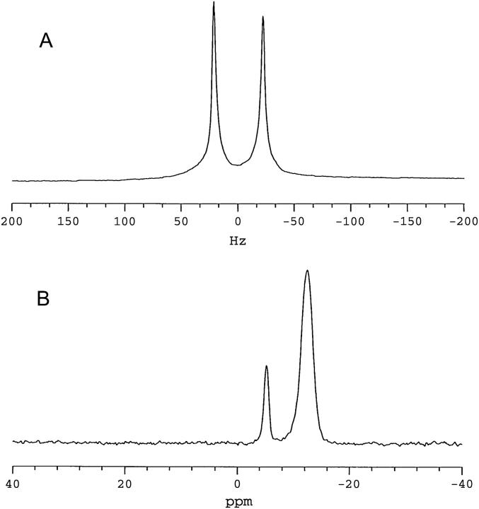
FIGURE 1.
NMR spectra (35°C) of magnetically aligned bilayers: DMPC/DHPC (q = 4.7, 25 wt % lipid) + 1 mol % DMPE-PEG 2000. (A) 2H NMR spectrum of HDO. The residual quadrupolar splitting of 44 Hz indicates magnetic alignment of bilayers. (B) 31P NMR spectrum showing resonances from DMPC (−12.45 ppm) and DHPC (−5.33 ppm), DMPC/DHPC intensity ratio of 4.7 ± 0.2. The position of the DMPC resonance is indicative of bilayers aligned with their normal to the plane of the bilayer oriented perpendicular to the direction of the magnetic field. The resonance from 1 mol % DMPE-PEG 2000 is not resolved.

