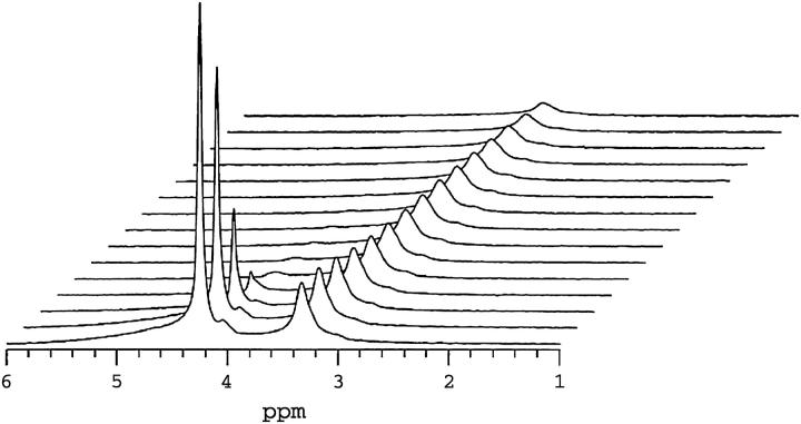
FIGURE 3.
1H NMR spectra (35°C) of magnetically aligned DMPC/DHPC (q = 4.7, 25 wt % lipid) + 1 mol % DMPE-PEG 2000 bilayers as a function of the field-gradient-pulse duration δ in the STE PFG NMR sequence. The two major resonances are assigned to HDO (4.2 ppm) and overlapping DPME-PEG 2000 ethoxy and DHPC choline methyl protons (3.3 ppm). Other lipid resonances are not visible due to their short transverse relaxation times relative to the spin-echo delay (10 ms) used in the acquisition of this spectrum. The gradient-pulse amplitude was 250 G cm−1, whereas τ2 equaled 10 ms and τ1 equaled 200 ms. Gradient pulse durations were, from front to back in units of ms, 0.1, 0.25, 0.50, 0.75, 1.0, 1.25, 1.5, 1.75, 2.0 2.5, 3.0, 3.5, 4.0, 5.0, and 6.0. The water resonance at 4.2 ppm decays rapidly due to water's fast diffusion. The combined DHPC choline methyl and DMPE-PEG 2000 headgroup resonance at 3.3 ppm decays far more slowly, as expected for bilayer intercalated species.

