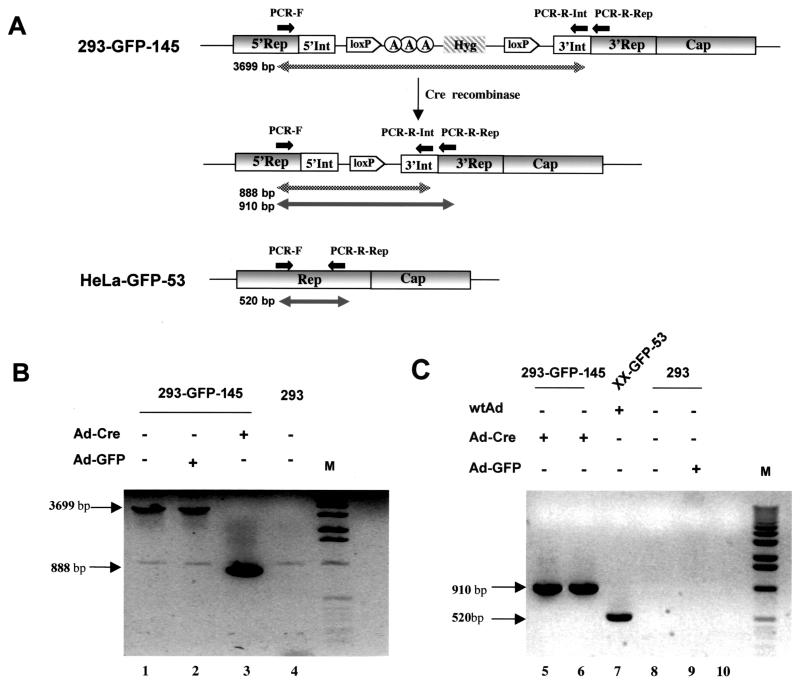
FIG. 8.
PCR analysis of AAV Rep gene structure before and after Ad-Cre and loxP-mediated DNA splicing. (A) Schematic illustration of the rep gene structure and expected sizes of PCR products. Primers PCR-F and PCR-R-Rep were located in the rep gene before and after, respectively, the inserted intron. Primer PCR-R-Int was located in the 3′ end of the intron. (B) Gel electrophoresis of PCR products with primers PCR-F and PCR-R-Int. Total cellular DNA was isolated from cell line 293-GFP-145 (lanes 1, 2, and 3) or control 293 cells (lane 4) with or without the indicated adenovirus infection. The DNA was subjected to PCR amplification and gel separation. (C) Gel electrophoresis of PCR products with primers PCR-F and PCR-R-Rep. Total cellular DNA (lanes 5, 7, 8, and 9) or episomal DNA (lane 6) was isolated from different cells with or without indicated adenovirus infection. Cell line XX-GFP-53 (lane 7) contain wild-type Rep sequence and yielded a 520-bp PCR product, whereas cell line 293-GFP-145 contains an inserted intron sequence and a loxP site (after Cre-mediated splicing) and yielded a 910-bp PCR product from both total cellular DNA (lane 5) and episomal DNA (lane 6).

