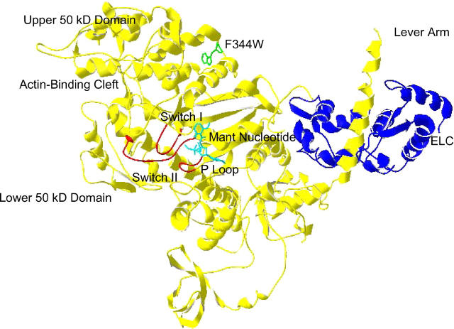
FIGURE 1.
Structure of F344W-MDE with the mant nucleotide (cyan) and the 344W residue shown as a ball-and-stick representation (green). Also highlighted are the essential light chain (ELC, blue) and the switch I, switch II, and P-loop (red). The chicken smooth-muscle myosin MDE structure (1BR4.pdb (2)) was superimposed with mant ADP coordinates (1LVK.pdb (4)), and a distance of 22.4 Å was measured between 344W and the mant moiety. This figure was prepared in Swiss PDB Viewer (GlaxoSmithKline).

