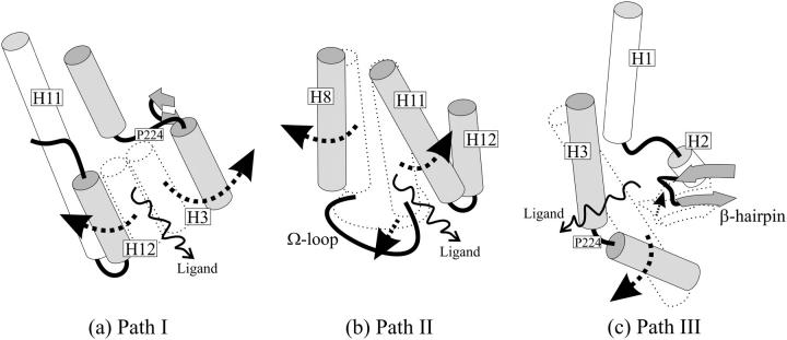
FIGURE 3.
Schematic representation of the three paths. (a) In Path I, H3 breaks down in two helices and H12 swings apart from it, forming the escape cavity. (b) The joint bending of H8 and the Ω-loop away from H11 allows ligand escape in Path II. (c) In Path III, the breakdown of H3 allows for the formation of a cavity between it and the β-hairpin through which ligands escape. Arrows indicate protein movements that from the bound sturcture (dotted) allow the ligands to escape.

