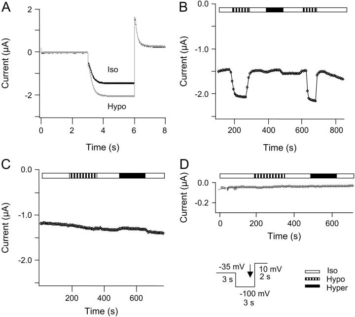
FIGURE 1.
Regulation of HCN2 channel current by changes in cell volume. HCN2 and AQP1 channels were expressed in X. laevis oocytes, and currents were activated by a step protocol, as indicated. Whole-cell currents were recorded at the end of the hyperpolarization step as indicated by the black arrow in the inset. The oocytes were superfused by isoosmotic extracellular solution followed by exposure to hypoosmotic extracellular solution and to hyperosmotic extracellular solution. (A) Current traces recorded in isoosomotic extracellular solution (black) and in hypoosmotic extracellular solution (shaded). For oocytes coexpressing HCN2 and AQP1 the mean increase in current after exposure to hypoosmotic extracellular solution was 30 ± 14% (n = 16). (B) The current depicted as a function of time after changes in extracellular osmolarity for the same experiment. (C) Time course of the whole-cell current in an oocyte expressing HCN2 channels without AQP1. The currents were recorded as described for B. The current was not significantly increased by exposure to hypoosmotic solution (1 ± 7%, n = 8, paired t-test, p = 0.9805) upon exposure to hypoosmotic extracellular solution. (D) Time course of whole-cell current in a native oocyte. The currents were recorded as described for A (n = 4).

