
FIGURE 4.
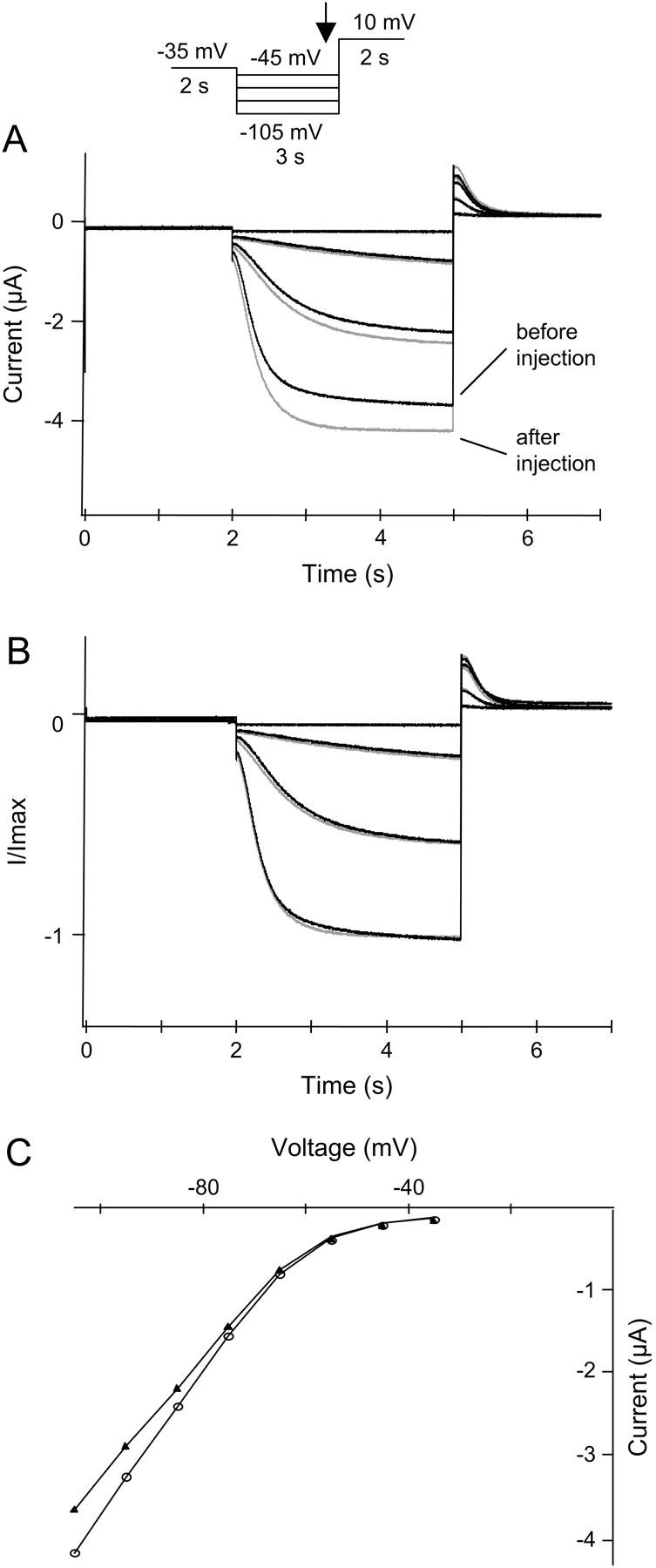
HCN2 currents are increased upon isoosmotic cell swelling. HCN2 channels were expressed in X. laevis oocytes and currents were activated by a step protocol as illustrated. Currents were measured in an isoosmotic extracellular solution before and during isoosmotic cell swelling. (A) HCN2 currents recorded before volume injection (black traces) and after injection of 50 nl intracellular solution (shaded traces), resulting in a current increase of 21 ± 4% (n = 4). (B) Normalized current traces from A. (C) The corresponding I-V curves (plotted as the current measured at the end of the hyperpolarization versus voltage). (▴) Currents recorded before injection. (○) Currents measured after injection of 50 nl intracellular solution.