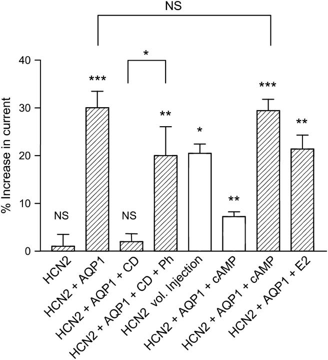
FIGURE 7.
Summary of the response of HCN2 channels to various stimuli. Relative responses as compared to controls are shown after different stimuli: (HCN2) For X. laevis oocytes expressing HCN2 and not AQP1, there was no significant increase in current after exposure to extracellular hypoosmotic solution (1 ± 7%, n = 8, paired t-test, p = 0.9805). (HCN2 + AQP1) For oocytes coexpressing HCN2 and AQP1, the current increased by 30 ± 14% after exposure to hypoosmotic extracellular solution. This increase is highly significant (n = 16, paired t-test, p = 0.0003). (HCN2 + AQP1 + CD) After exposure to 10 μM CD for t > 3 h before the experiments, there was no significant response to hypoosmotic volume changes (2 ± 7%, n = 9, paired t-test, p = 0.1920). (HCN2 + AQP1 + CD + Ph) The response could be at least partially rescued by adding 25 μM phalloidin to the 10 μM CD solution (20 ± 16%, n = 7, paired t-test, p = 0.0097). (HCN2 vol. Injection) Isoosmotic cell swelling of HCN2-expressing X. laevis oocytes was induced by injection of 50 nl isoosmotic solution. Injection resulted in a significant current increase by 21 ± 4% (n = 4, paired t-test, p = 0.0138). (HCN2 + AQP1 + cAMP) Increased cAMP resulted in an enhancement of HCN2 current of 7 ± 2% (n = 5, paired t-test, p = 0.001, open bar). Cell swelling in the presence of cAMP significantly increased HCN2 current by 29 ± 5% (n = 5, paired t-test, p < 0.0001) compared to isoosmotic media without cAMP. (HCN2 + AQP1 + E2) Coexpression of HCN2, AQP1, and KCNE2 resulted in a threefold current increase compared to oocytes expressing HCN2 and AQP1 alone (data not shown). The relative current increase upon exposure to hypoosmotic solution for HCN2, AQP1, KCNE2-expressing oocytes was 21 ± 6% (n = 4, paired t-test, p = 0.0069).

