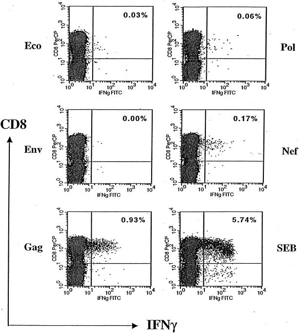
FIG. 2.
Quantifying HIV-1-specific CD8+ T cells by intracellular cytokine staining. Aliquots of PBMC from one HIV-1-infected individual were stimulated by recombinant vaccinia virus expressing negative control Eco, HIV-1 antigens (Env, Gag, Pol, and Nef), and positive control SEB first, followed by staining with anti-CD3, -CD4, -CD8, and -IFN-γ antibodies. The antigen-specific CD8+ T cells were enumerated and expressed as a percentage, shown on the upper right quadrant of each plot. FITC, fluorescein isothiocyanate.

