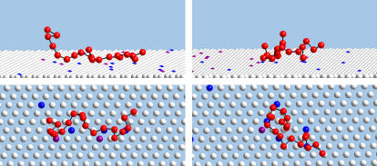
FIGURE 2.
Side and top views of two, rather arbitrary, simulation snapshots (left and right), of a polyelectrolyte interacting with a weakly charged fluid membrane (1% PIP2, and 1% PS). For visual clarity only a section of the membrane is shown, and polymer segments and lipid headgroups are depicted as small spheres, (recall, however, that short range repulsions keep these segments at distance ≥ d). PIP2 and PS lipids are represented by blue and purple spheres, respectively. Note the localization of the charged lipids in the vicinity of the polymer.

