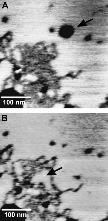
FIGURE 2.
AFM topography images of peptide domains in a DPPC bilayer before (A) and after (B) peptide extraction. The arrow in A points to a place where lipids have been extracted. In B, the bilayer has healed. The arrow in B points to the space left by the extracted peptides, which has been filled by lipids.

