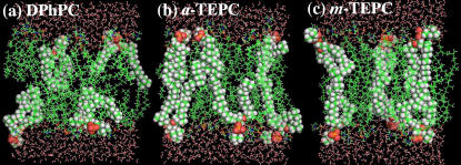
FIGURE 5.
Snapshots (20 Å slab) of the membrane systems during the MD simulation. (a) DPhPC, (b) a-TEPC, and (c) m-TEPC. Phosphorus atoms are yellow, nitrogens are blue, oxygens are red, carbons are green, and hydrogens are white. Atoms in several selected lipid molecules are represented by spheres to show typical lipid conformations clearly.

