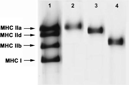
FIGURE 1.
Silver-stained polyacrylamide gel electrophoresis pattern of myosin heavy-chain (MHC) isoforms from rabbit skeletal muscle fibers. Lane 1 represents a mixture of extracts from rabbit gastrocnemius and diaphragm muscles and serves as marker for MHC-IIb, MHC-IId, MHC-IIa, and MHC-I as indicated. The other lanes represent single fibers for which mechanical properties were measured: lane 2, type IIA (EDL); lane 3, type IID (adductor magnus); lane 4, type IIB (adductor magnus).

