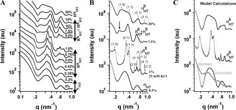
FIGURE 3.
Small angle synchrotron x-ray diffraction scans of MTs with 20 k PEO. (A) The scattering patterns continuously evolve as the 20 k PEO concentration is increased from 0% (wt/wt) to 20%. All peaks, oscillations, and minima of the scattering can be accounted for by three structures: a nematic of single MTs (NMT), bundles with rectangular symmetry ( ), and bundles with hexagonal symmetry (
 ). (B) Up to nine peaks of the rectangular lattice and four peaks of the hexagonal lattice are visible, all of which can be indexed. The (1 0) rectangular peak is often difficult to discern but is more prominent with 25 mM added KCl (
 ). Coexistence between bundles with rectangular and hexagonal symmetry is evident in many scans (
 arrow indicates hexagonal (1 0) peak. All other peaks are from rectangular bundles, indices not shown). (C) Model calculations of the scattering from isolated MTs (unbuckled), unbundled, distorted MTs (buckled), bundles of distorted MTs with rectangular symmetry (
 ), and bundles of undeformed MTs with hexagonal symmetry (
 ). See text for modeling details.

