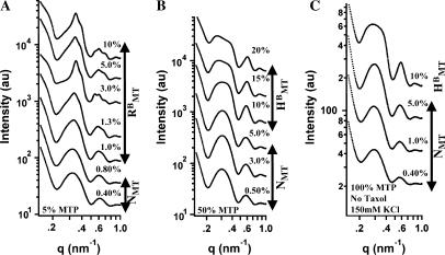
FIGURE 6.
Small angle synchrotron x-ray diffraction scans of 20 k PEO with MTs, polymerized in the presence of partially purified MTP, which is ∼30% MT-associated protein and ∼70% tubulin. (A) MTs polymerized with 5% MTP form rectangular bundles but only with higher concentrations of 20 k PEO. (B) With 50% MTP hexagonal bundles are present for high 20 k PEO concentrations and rectangular bundles are not observed. (C) 100% MTP with no taxol and 150 mM KCl also display no rectangular bundles.

