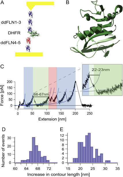
FIGURE 1.
(A) Schematic representation of the Ddfilamin-DHFR protein construct, stretched between a gold surface and a gold-coated cantilever tip. (B) Proposed structure of the intermediate state (dark green). The C-terminal part shown in light green unfolds during the first unfolding event. (C) Typical force versus extension curve. The unfolding pattern of the Ddfilamin domains is shown in blue (and in red for domain 4), whereas the unfolding event of DHFR is shown in green. The inset shows a magnification in which the intermediate state is clearly visible. (D) Increase in contour length for the complete unfolding of DHFR. (E) Increase in contour length for the unfolding of DHFR from the native state to the intermediate state.

