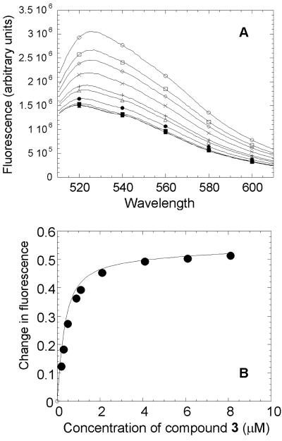
FIG. 3.
Determination of binding affinity of compound 3 for E. coli MurB. (A) Fluorescence spectra of FAD bound to E. coli MurB in the absence and presence of compound 3. Each curve (described in order from top to bottom) represents one wavelength scan (excitation at 460 nm) of E. coli MurB at 1 μM alone or with 1, 2, 3, 4, 5, 6, 7, or 8 μM compound 3. (B) Binding isotherm for the interactions of compound 3 with E. coli MurB. Compound 3 binds E. coli MurB tightly with a Kd of 260 nM.

