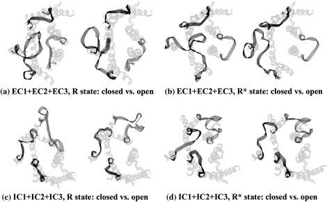
FIGURE 4.
Pairs of the most closed (left structures) and most opened (right structures) low-energy conformers of the extracellular (panels a and b) and the intracellular (panels c and d) for the R and R* states of rhodopsin (left and right panels, respectively). The loop structures are shown as five-line ribbons in dark gray. TM region of rhodopsin is shown as five-line ribbon in light gray. Loops are shown in clockwise order from EC2 to EC1 to EC3 (a and b) or from IC1 to IC2 to IC3 (c and d) starting from the lower left corner of the figure. Views are from the extracellular (a and b) and intracellular (c and d) sides of the membrane, respectively.

