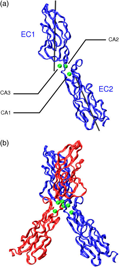
FIGURE 1.
Ribbon diagrams of EC1-2 fragments of E-cadherin. (a) Monomer, showing the three calcium ions (in green) bound at the domain junction and the axes used to characterize the relative position of the two domains. (b) cis-dimer of EC1-2 (PDB file 1FF5, (11)). The images in Figs. 1 and 4 were prepared using VMD (33).

